Describe and State the Uses of Coverslip
Depending on the sample a slide and cover slip is used at times to flatten and contain the sample. HANGING DROP PREPARATION STEP 3.
Microscope Cover Slips Explained
The purpose of coverslipping is.

. Protect tissues from scratching being removed from the slide. Difficult to see with the light microscope. If your specimen is a littler larger you can use tweezers or whatever tool that will help you place the specimen in the center of the slide without damaging it killing it or contaminating it.
This lab will help you practice the following skills. The sample is preserved or prepared with a medium stain by placing a cover slip on top of the sample on the slide. However imaging at higher resolutions from 40X through 100X objectives must be imaged through a very thin layer of glass no thicker than a standard coverslip.
This creates a small well with a glass bottom onto which cells can be grown. Microscopy imaging with an objective lens from 1X through 40X at lower resolutions is extremely flexible and can be performed on a very wide variety of samples. Coverslips chambers and working distance.
This video explains step-by-step how to mount a coverslip properly for cellular imaging as illustrated in steps 1-4 of the below protocol. When viewing sections or smears typically biological in nature they must be fixed to a slide. The coverslip protects both your specimen and the objective of the microscope from damage.
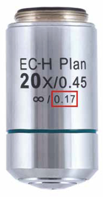
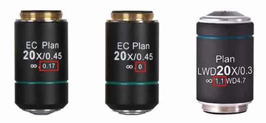
Coverslip thickness is less important when using objective lenses that have a numerical aperture NA of 04 or lower which on most microscopes would only be lenses between 1x-10x. Of the coverslip and add the substance to the opposite edge of the coverslip one drop at a time. 1 12 thickness.
The first step is to use the Q-tip to swab your sample and apply it to the center of the microscope slide. Bacterial cells in the living state were first observed by Leeuwenhoek in drops of fluid. Up to 24 cash back use substance X.
Using a light microscope 2. Remove coverslip containing the sample from the buffer. Examine the preparation under low power objective lens with reduced light and close the diaphragm of the microscope.
Use only enough mountant to fill the space on the coverslipslide and not excess and this assessment comes with experience. Too little mounting media will cause air bubble at the edges of coverslip and one will be tempted to press down on the coverslip to. We describe an emulsion-coated coverslip autoradiographic technique for large 50 x 50 mm sections of monkey or human brain.
To prepare a wet mount using a flat slide or a depression slide. Once the liquid has been added to the slide a coverslip is placed on top and the specimen is ready for examination under the microscope. Lift the preparation and quickly turn the hanging drop preparation coverslip up so that the culture drop is suspended in the concavity of Depression slide.
The fixing of a sample refers to the process of attaching cells to a slide. In this report we offer an improved method using Parafilm as a dry mount adhesive for the preparation of special. The coverslip protects the.
In addition to providing better optical qualities the glass-bottomed well has a very small volume and therefore is ideal for conserving limited reagents such as those used in immunocyto- chemistry. A small square of clear glass or plastic a coverslip is placed on top of the liquid to minimize evaporation and protect the microscope lens from exposure to the sample. Ing a coverslip under a hole bored in the bottom of a plastic petri dish.
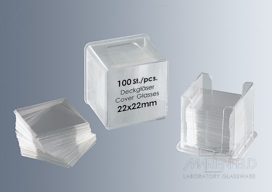
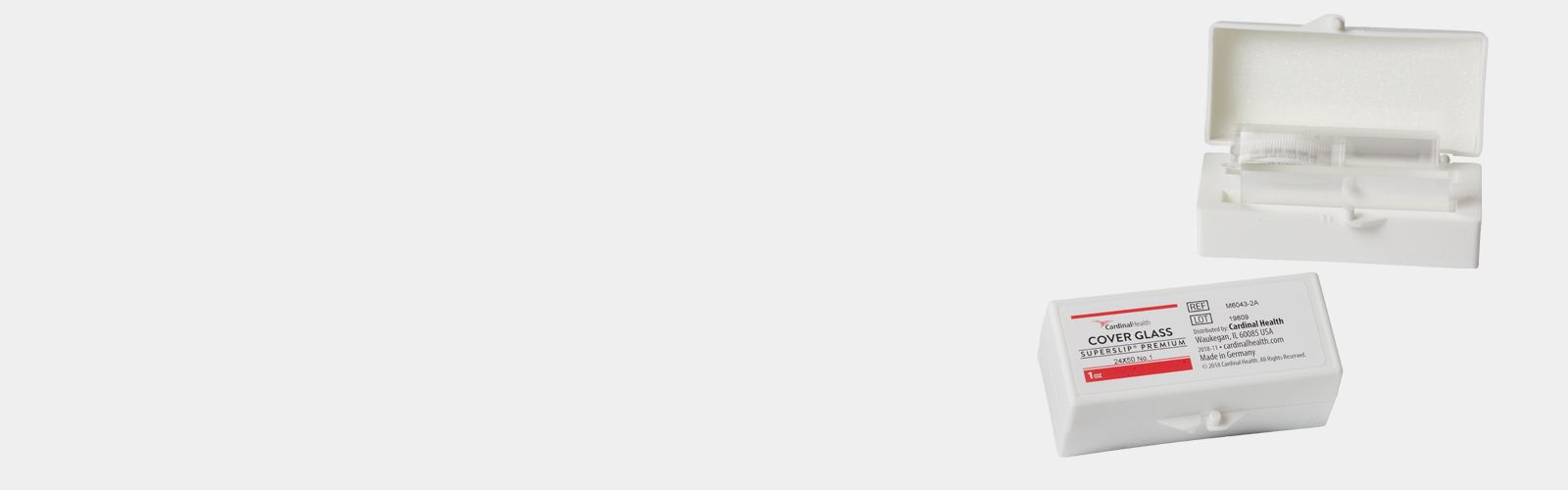
The graph shown used a. Commercial sealants laboratory preparations clear nail polish or other adhesives can be used. Coverslips are available in various shapes sizes and thicknesses.
For such applications laboratories often prepare special dishes which are made by affixing a glass coverslip beneath a hole in a plastic petri dish bottom. Fixing - preserve specimens in as near natural state as possible Sectioning - dehydrated with alcohols placed in a waxresin to form hard block - sliced very thinly by a microtome Staining - multiple stains for different structures Mounting - secured to slide coverslip on top sealed to create permanent slide. HANGING DROP PREPARATION STEP 4.
As magnification 2x or higher and NA increases the loss from having an incorrect thickness coverslip can become significant. Place a clean dry 24 mm 50 mm glass coverslip onto the silicone gasket and press gently to seal. Describe a procedure that could be used to add this substance to the cells on the slide without removing the coverslip.
Keep tissue as well as stain from drying out improve the refractive index prtect stain from fading. The purpose of this lab is to use a compound light microscope to examine several specimens and to make wet mounts. Add more drops as the.
For many applications cells or tissue must be cultured on an optical surface of high quality. The method was adequate and even desirable for certain observations but the. The second method of preparing specimens for light microscopy is fixation.
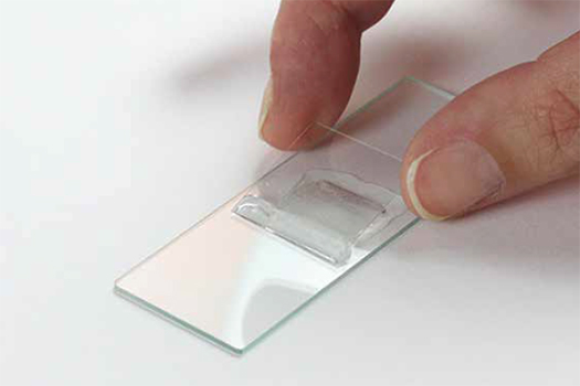
A coverslip in Biology is a small rectangle of glass that is mounted on a slide to be viewed under a microscope. It also protects permanent slide. Sometimes the liquid used is simply water but often stains are added to enhance contrast.
The thickness can affect the resolution and intensity of the image. A new coverslip use a piece of adhesive tape to clean lint and debris from the gasket surface. The technique uses adhesive-backed teflon-reinforced aluminum foil as a flexible hinge that allows the coverslip to swing away from the slide so that the emulsion and tissue can be processed independently.
Coverslips can be glued to the slide to preserve the specimen slow drying and prevent contamination. Apply a small amount of mounting medium to the surface of the slide. Moisture will ruin the seal around the edge of the lens unless a lens is specially made to focus through water or oil.
Wet mounts were commonly used by Pasteur Lister and many of the early workers in the field of microbiology. Introduction to Microscopy Purpose. Try to use an amount that will just fill the space under the coverslip.
Place gasketed coverslips individually into glass petri. This protects objective lens of microscope from getting stain from a wet mount. Identify the type of organelle in cell A that.
Creating wet mount slides to learn how handle and stain fresh tissues. Coverslip is not placed on a microscope but on the stained specimen on slide.
Microscope Cover Slips Explained

File Coverslip Graphic Png Wikimedia Commons

Indstb003ueqm8w Capitolbrand M34532424 Glass Microscope Slide Cover Slip 24mm Length 24mm Width 1 Thick Box Of 100

Coverslips Cover Glass And Film

Microscopy 5 Reasons Coverslips Are Important For High Quality Imaging

Coverslips Cover Glass And Film
Microscope Cover Slips Explained

Mounting Tissue Sections National Diagnostics
Cover Glasses Australian Scientific Microscope Coverslip

Why Do We Add Coverslip On A Slide While Watching Under The Microscope Brainly In

Microscopy 5 Reasons Coverslips Are Important For High Quality Imaging

Microscopy 5 Reasons Coverslips Are Important For High Quality Imaging
Exploded View Of A Sac Growth Chamber Showing A Microscope Slide With Download Scientific Diagram
Coverslips Cover Glass And Film

Coverslips Cover Glass And Film
Coverslips Cover Glass And Film

Micromachines Free Full Text Micromirror Embedded Coverslip Assembly For Bidirectional Microscopic Imaging Html

Coverslips Cover Glass And Film

Micromachines Free Full Text Micromirror Embedded Coverslip Assembly For Bidirectional Microscopic Imaging Html

Comments
Post a Comment